
Image bank
Dr John Walker
Dr John Walker is a University of Sydney alumni who worked at the Sydney School of Public Health and Westmead Hospital as a Senior Lecturer in Parasitology for 38 years. His work focused on diagnostic parasitology and epidemiological surveys of gastrointestinal parasites, malaria and filariasis. A major emphasis of his latter work was providing malaria reference laboratory services and maintenance of a register of malaria cases imported into NSW. Dr Walker retired in 2005 but has retained his interest in parasitology.
Image bank samples
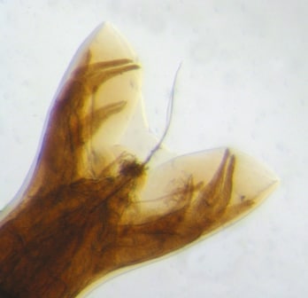
Necator americanus, adult male, bursa from human faeces tissue.
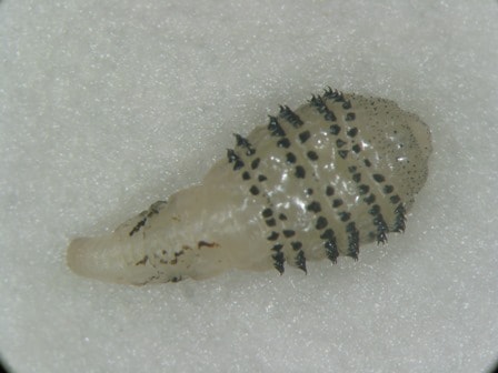
Dermatobia hominis, larva from human subcutaneous lesion tissue.

Fasciola hepatica, section of adult worm from human cervical lymph node tissue.
Trichuris trichiura, male cloacal region showing spicule from human faeces tissue.

Echinococcus granulosus, Hydatid cyst, protoscolices from human liver tissue.

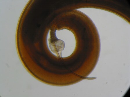
Strongyloides stercoralis, third stage filariform larva from human sputum tissue.

Ascaris lumbricoides, immature egg from human faeces tissue.

Diphyllobothrium latum, coracidium released from egg in human faecal smear.