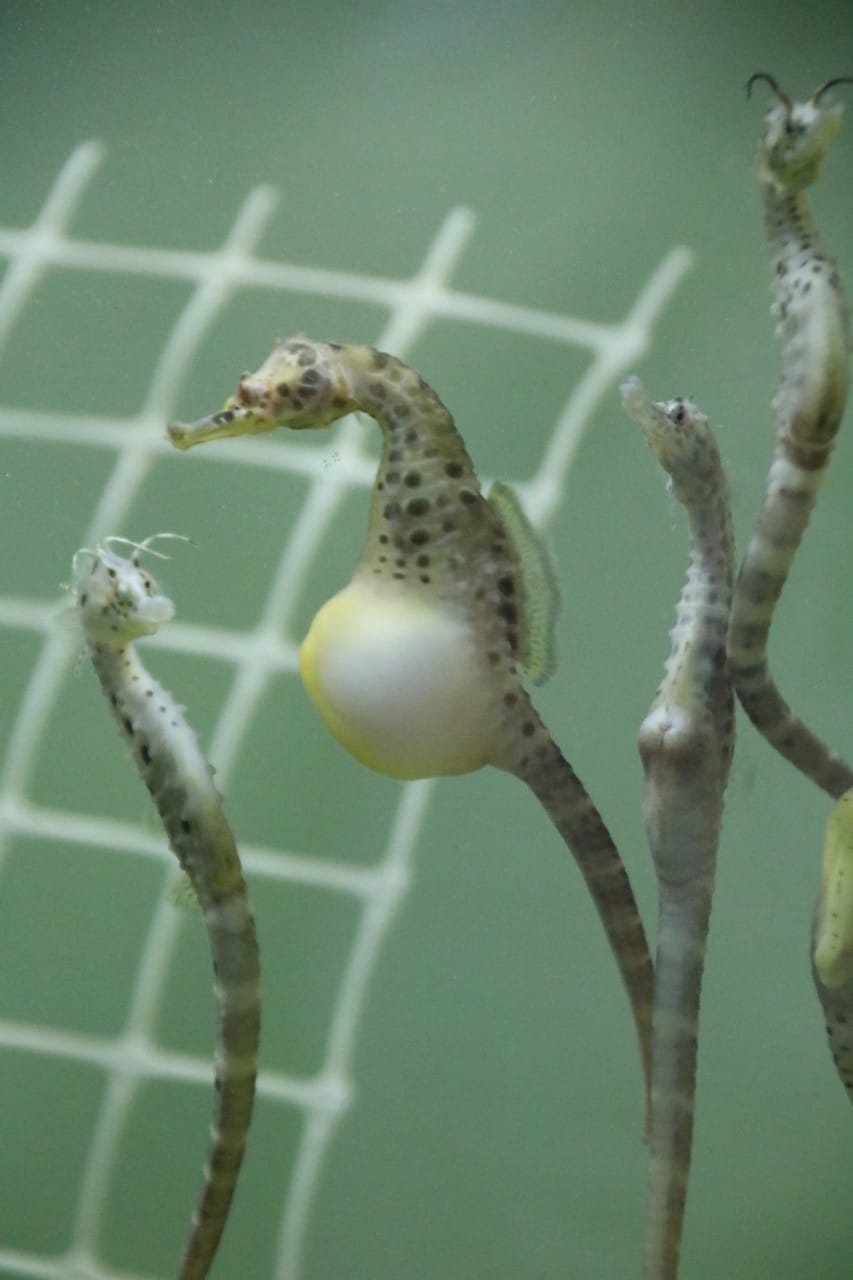
Male seahorses develop placentas to support their growing babies

By viewing the seahorse pouch under the microscope at various stages of pregnancy, researchers found that small blood vessels grow within the pouch. Image credit: Jacquie Herbert.
Supplying oxygen to their growing offspring and removing carbon dioxide is a major challenge for every pregnant animal. Humans deal with this problem by developing a placenta, but in seahorses — where the male, not the female, gestates and gives birth to the young — exactly how it worked hasn’t always been so clear.
Male seahorses incubate their embryos inside a pouch, and until now it was unclear how the embryos “breathe” inside this closed structure. Our new study, published in the journal Placenta, examines how pregnant male seahorses (Hippocampus abdominalis) provide oxygen supply and carbon dioxide removal to their embryos.
We examined male seahorse pouches under the microscope at different stages of pregnancy, and found they develop complex placental structures over time — in similar ways to human pregnancy.
A pregnant dad gestating up to 1,000 babies
Male pregnancy is rare, only occurring in a group of fish that includes seahorses, seadragons, pipehorses and pipefishes.
Pot-bellied seahorse males have a specialised enclosed structure on their tail. This organ is called the brood pouch, in which the embryos develop.
The female deposits eggs into the male’s pouch after a mating dance and pregnancy lasts about 30 days.
While inside the pouch, the male supplies nutrients to his developing embryos, before giving birth to up to 1,000 babies.
Embryonic development requires oxygen, and the oxygen demand increases as the embryo grows. So too does the need to get rid of the resulting carbon dioxide efficiently. This presents a problem for the pregnant male seahorse.
Enter the placenta
In egg-laying animals — such as birds, monotremes, certain reptiles and fishes — the growing embryo accesses oxygen and gets rid of carbon dioxide through pores in the egg shell.
For animals that give birth to live young, a different solution is required. Pregnant humans develop a placenta, a complex organ connecting the mother to her developing baby, which allows an efficient exchange of oxygen and carbon dioxide (it also gets nutrients to the baby, and removes waste, via the bloodstream).
Placentae are filled with many small blood vessels and often there is a thinning of the tissue layers that separate the parent’s and baby’s blood circulations. This improves the efficiency of oxygen and nutrient delivery to the fetus.
Surprisingly, the placenta is not unique to mammals.
Some sharks, like the Australian sharpnose shark (Rhizoprionodon taylori) develop a placenta with an umbilical cord joining the mother to her babies during pregnancy. Many live-bearing lizards form a placenta (including very complex ones) to provide respiratory gases and some nutrients to their developing embryos.
Our previous research identified genes that allow the seahorse father to provide for the developing embryos while inside his pouch.
Our new study shows that during pregnancy the pouch undergoes many changes similar to those seen in mammalian pregnancy. We focused on examining the brood pouch of male seahorses during pregnancy to determine exactly how they provide oxygen to their developing embryos.
What we found
By viewing the seahorse pouch under the microscope at various stages of pregnancy, we found that small blood vessels grow within the pouch, particularly towards the end of pregnancy. This is when the baby seahorses (called fry) require the most oxygen.
The distance between the father’s blood supply and the embryos also decreases dramatically as the pregnancy goes on. These changes improve the efficiency of transport between the father and the embryos.
Interestingly, many of the changes that occur in the seahorse pouch during pregnancy are similar to those that occur in the uterus during mammalian pregnancy.
We have only scratched the surface of understanding the function of the seahorse placenta during pregnancy.
There is still much to learn about how these fathers protect and nourish their babies during pregnancy — but our work shows the morphological changes to seahorse brood pouches have a lot in common with the development of mammalian placentae.
This article was first published on The Conversation and written by Dr Camilla Whittington from University of Sydney and Dr Jessica Suzanne Dudley from Macquarie University.

