Digital diagnostic imaging services
University Veterinary Teaching Hospital Sydney (UVTHS) is a fully digital Radiology Department comprising of the following services:
- Picture Archive and Communications System (PACS)
- Digital Radiography (DR)
- Ultrasound (US)
- Computed Tomography (CT)
- Magnetic Resonance Imaging (MRI) systems.
- Fluoroscopy
The benefits of Digital Imaging are numerous as the system allows easy imaging access to veterinarians, researchers and students. Clients are able to review pet’s images with their veterinarian during consultation, making the diagnosis and treatment process more efficient. A digital copy containing pet’s images and radiology reports can be made available to clinicians on request.
All imaging modalities are DICOM 3.0 compliant.

General radiography
General radiology services include all routine diagnostic X-ray procedures plus Image Intensified Video-fluoroscopy for myelography, excretory urogram, retrograde vaginourethrogram and other real time procedures.
Digital radiography (DR)
DR technology has enabled the UVTHS to enhance standard X-ray images through the use of optimised radiographic algorithms. Studies have been further improved with the use of High Detail and High Speed cassettes depending upon the needs of examination. High detail bone imaging or fast chest radiographs are easily obtained with DR imaging.
Orthopaedic Templating for surgical planning is another advantage of digital radiology. Our surgeons can plan hip or knee pin surgery in advance of the actual procedure to insure the best patient’s outcome possible.

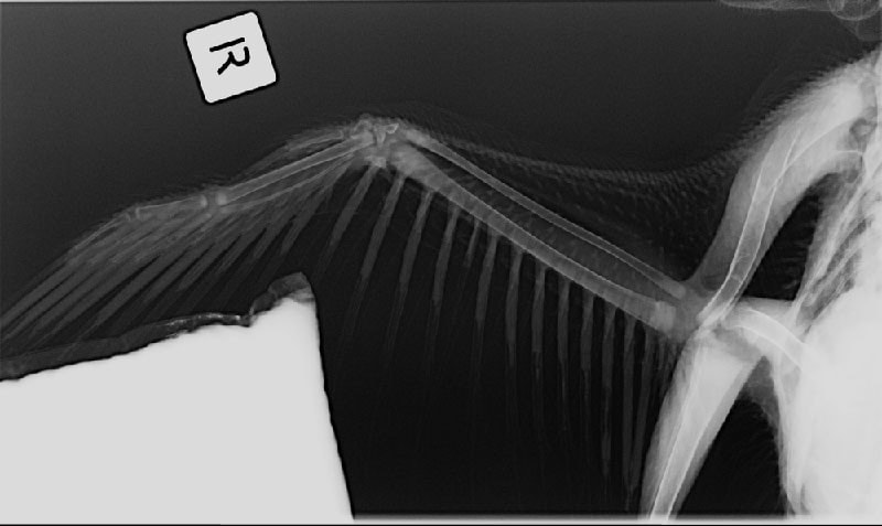
Ibis wing

Cat abdomen

Cat asthma
Available examinations
- Abdominal, cardiac and vascular studies performed using B-mode
- Colour flow Doppler, pulsed wave Doppler and continuous wave Doppler
- Harmonic imaging and contrast ultrasound
Ultrasound guided procedures
- Fine needle aspirates and biopsies
- Therapeutic or diagnostic abdominocentesis
- Thoracocentesis or pericardiocentesis

Pancreas

Dog kidney

Bladder lesion
Computed tomography (CT)
CT imaging provides the UVTHS with high resolution cross sectional imaging of brain, spine, chest, abdominal and limb examinations.
CT imaging strength lies in its ability to image quickly for chest and abdominal studies so that images are not affected by the pet’s breathing. Exquisite bone detail is easily obtained for skull, sinuses, spine or limb studies.
The digital images can be rebuilt and viewed in a 3D volume movie providing additional information to veterinarians. The CT is a high end system used worldwide for both veterinary and human imaging.

Dog chest

Dog nose

3D dog pelvis
Magnetic resonance imaging (MRI)
MRI strength is seen in its ability to image soft tissues structures such as brain, eyes, pituitary gland, spinal cord, nerves, muscles, tendons and can differentiate normal tissue from tumours and various other disease processes.
MRI of the brain is often used to evaluate and define tumour types, infection, seizure disorders, strokes, hydrocephalus and other damage as a result of trauma. MRI of the spine is used for diagnoses of spinal cord tumours or syrinx, herniated disks, spinal cord compression and cord inflammation. Knee and hip MRI can be used to investigate tendon or ligament tears, bone or muscle tumours and inflammation.
The MRI 0.25 tesla open system is designed especially for small animals.

Dog brain

Dog lumbosacral spine

Dog left stifle

