About us
The Image X Institute is a centre for innovation in radiation therapy and cancer imaging technologies.
Our mission is to improve lives by inventing and advancing new ways to image and treat disease. We’re working on new technologies for medical imaging and targeted radiation therapy.
We engage with industry, hospitals, international collaborators and universities to forge relationships that help take our projects from lab bench to patient bedside.
Image X is a part of the Faculty of Medicine and Health at the University of Sydney. Explore more research within our faculty.
– View current Opportunities to work or study with us.
– Explore our research
– Meet our staff
– Follow us on twitter.
Latest News
April 2024
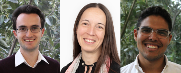
Trio of Grant Successes
Congratulations to Owen Dillon, Hilary Byrne and Nicholas Hindley (picture L-R), all of whom have been successful in recent funding rounds.
Owen Dillon: Creating the Next Generation in Radiotherapy Imaging with Carbon Nanotube Sources and Photon Counting Detectors. CCNSW Project Grant
Read more: Cancer Council NSW Announcement
Dr Hilary Byrne: How are you breathing today? Embedding functional imaging throughout radiation therapy to improve lung cancer patient outcomes. Cancer Australia ECR PdCCRS grant
Dr Nicholas Hindley: From relativity to respiration: How ideas from Einstein’s general theory enable adaptative radiation therapy for lung cancer patients. Cancer Australia Category A ECR PdCCRS grant
Read More: Cancer Australia Announcement
Jan 2024
Patent Awarded to David Waddington
Congratulations to David Waddington, who has been awarded a patent for “Systems and methods for susceptibility contrast imaging of nanoparticles at low magnetic fields”. In 2020, David published in Science Advances showing that iron-oxide nanoparticles could revolutionise the contrast possibilities for MRI, especially in low-field, portable setups.
Now, this patent, co-invented with collaborators Matthew Rosen from Massachusetts General Hospital and Zdenka Kuncic from University of Sydney, safeguards the game-changing technique David introduced in that paper.
Given the rapid expansion of the portable MRI market segment, we’re excited for the potential licensing opportunities for this patent. Watch this space!
Read the paper here: https://www.science.org/doi/10.1126/sciadv.abb0998

Nov 2023
EVENT: Research Showcase
You’re invited to join us on Thursday November 9 for an afternoon showcase of the incredible research happening here at Image X. Following the showcase, we are honoured to host a presentation from Professor Laurence Court from the MD Anderson Cancer Centre, University of Texas.
When: Thursday November 9th, from 2:30pm
Where: Seminar Room 422, Level 4, Biomedical Building 1 Central Avenue Eveleigh, NSW 2015
RSVP/More Info: register for free via Eventbrite by Nov 8th: Register + More Info

Contact us
Phone: +61 2 8627 1106
Email: image-x.contact@sydney.edu.au
Social media: Facebook | Twitter
Level 2, Biomedical Building (C81)
1 Central Ave
Australian Technology Park
Eveleigh NSW 2015
University Internal Mailing
Suite 201, C81-Biomedical Building,
University of Sydney, NSW 2006